Funakoshi

Funakoshi is an expert in research reagents such as dyes and instruments for life science researchers
Funakoshi Co., Ltd. has established itself as a leading distributor of research reagents and instruments for life science researchers since its inception. Their corporate mission revolves around providing high-quality products to a diverse customer base through their extensive domestic and international network.
They offer a wide range of products, including live imaging dyes, cell culture goods, natural compounds, and other related items.
Filter products

LipiDye II
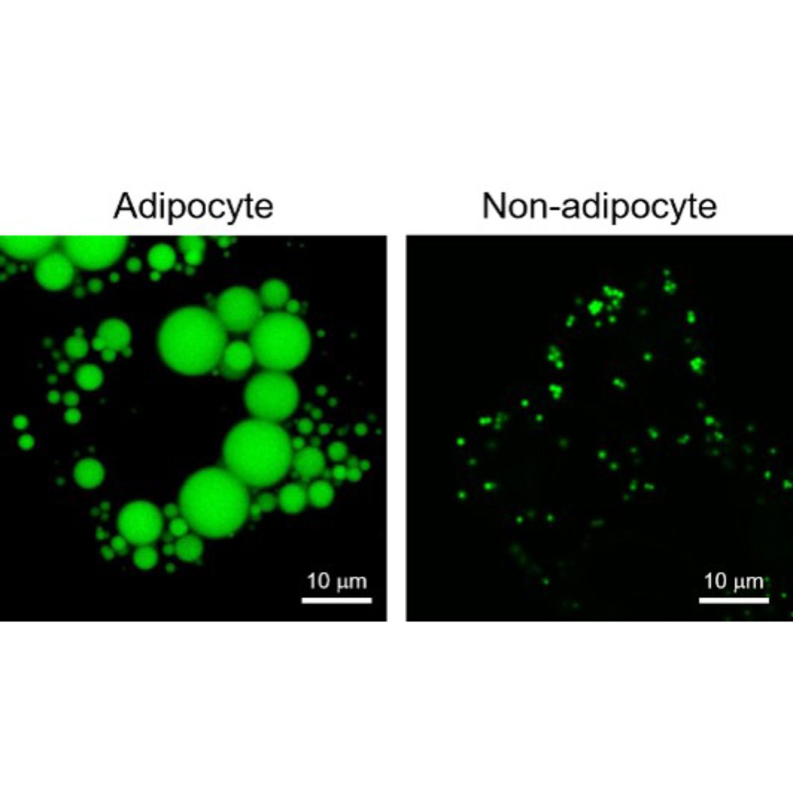
LipiDye II is an advanced fluorescent dye specifically designed for detecting Lipid Droplets (LDs) with exceptional sensitivity, minimal cytotoxicity, and remarkable photostability. This dye surpasses previous options by enabling the detection of even the smallest LDs (less than 1µm) and facilitating long-term time-lapse imaging, including Z-stack imaging. Through the application of this innovative dye, researchers can successfully observe the dynamic processes of LD biosynthesis, degradation, and movement within live cells.
Migrastatin
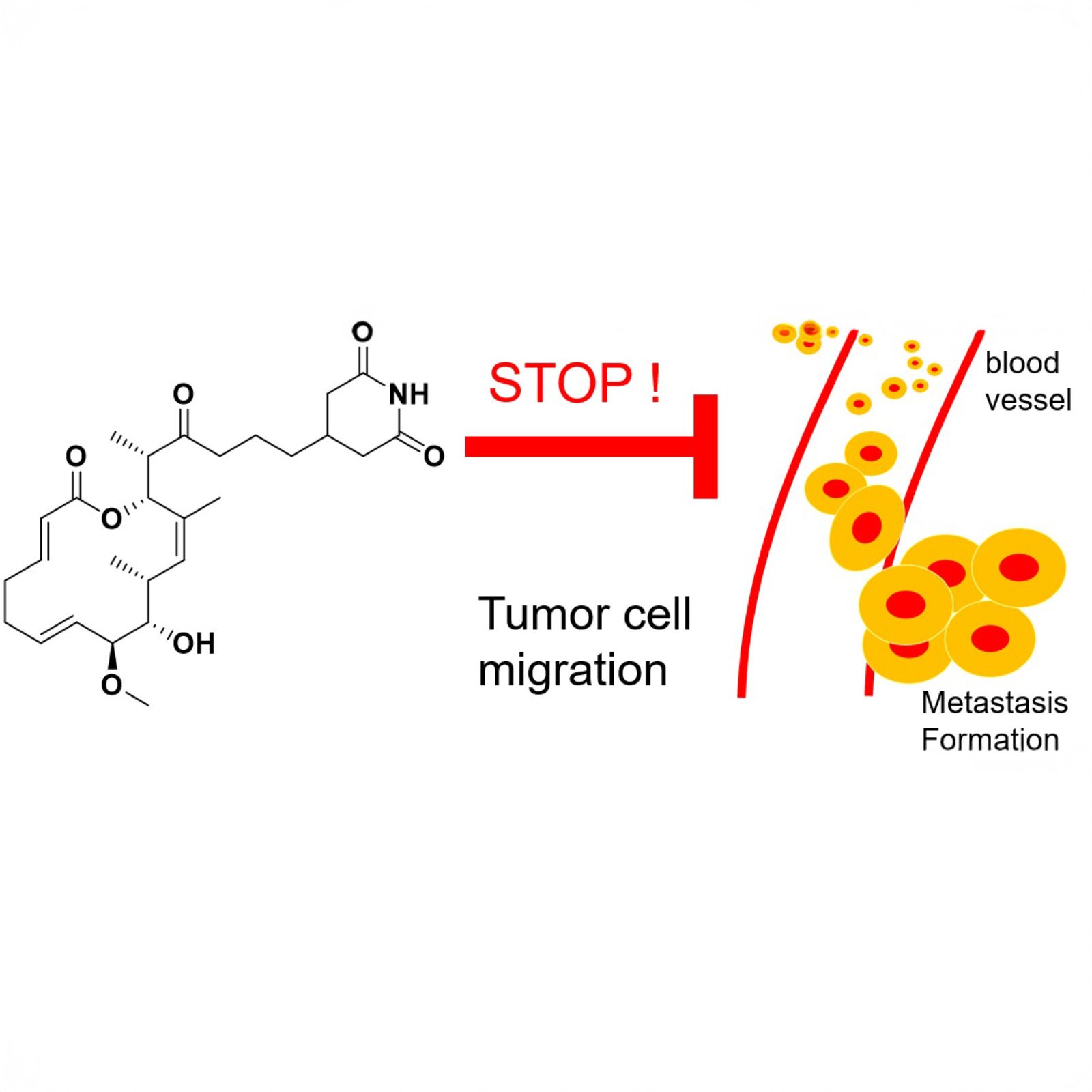
Migrastatin is a natural compound isolated from actinomycetes, initially reported in 2000, with extensive research conducted on its mechanism of action and analogues. It has demonstrated significant potential as an anti-cancer agent, specifically in inhibiting cancer cell migration and invasion. Through its interference with molecular pathways involved in cell migration, migrastatin may prevent the metastasis of cancer cells. Its structure-activity relationship has been explored, leading to the development of analogues with improved potency and specificity. The continued investigation of migrastatin and its analogues holds promise for the development of novel anti-cancer therapies and other medical applications.

NucleoSeeing
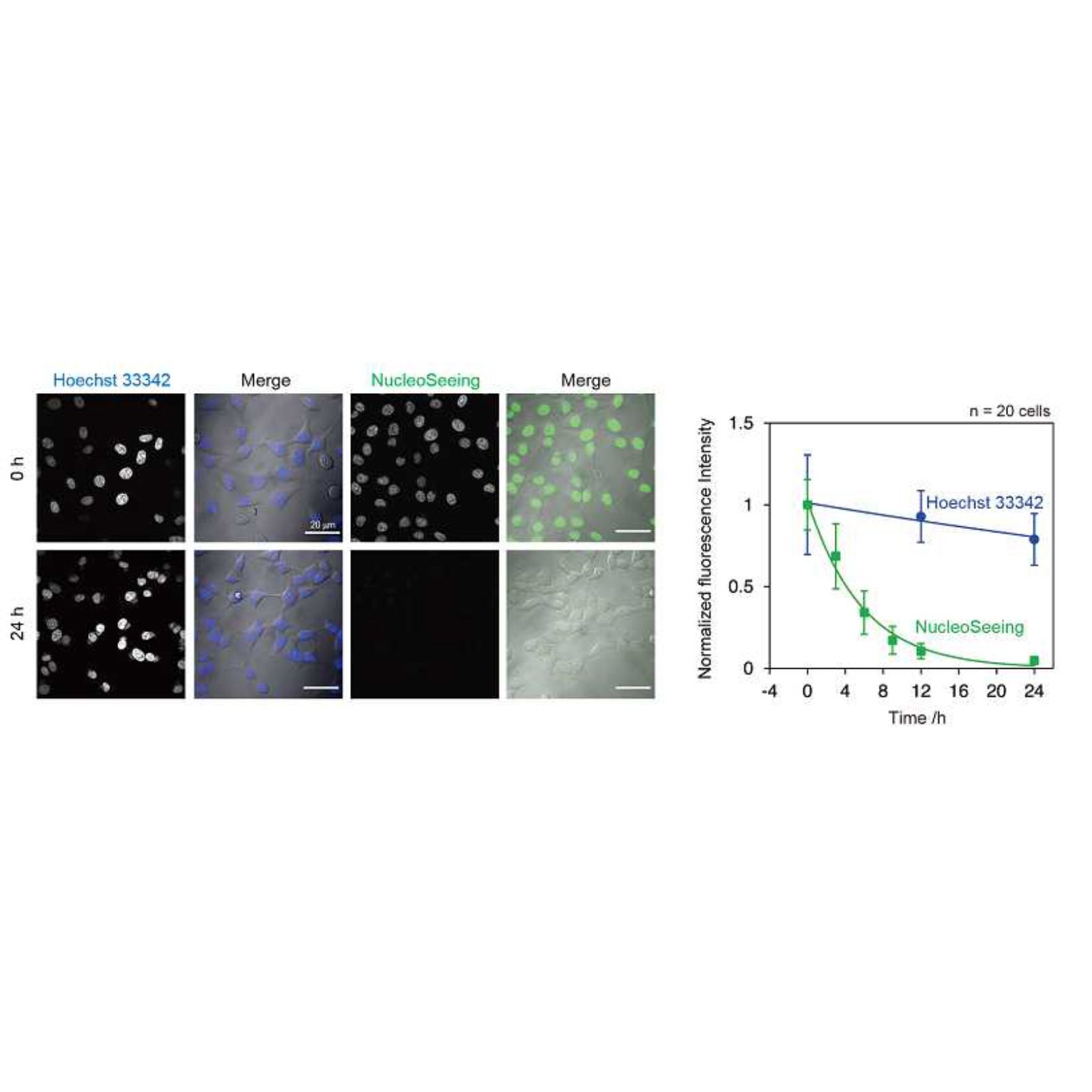
NucleoSeeing is an innovative live cell imaging probe that specifically binds to DNA, resulting in the emission of green fluorescence. This versatile product is not limited to animal cells or tissues but can also be utilized in Arabidopsis thaliana's Guard cells with a high signal-to-noise ratio. Additionally, NucleoSeeing serves as a pH sensor within the nucleus.
This commercially available product has been developed under a license from the Nagoya Institute of Technology. Comprising a green fluorescent dye and a DNA binding tag, NucleoSeeing remains in a folded conformation and does not emit fluorescence when not bound to DNA. However, upon binding to DNA, its conformation changes, leading to the emission of green fluorescence.
NucleoSeeing offers several advantages, including a high signal-to-noise ratio, excitation/emission wavelengths of 488 nm/520 nm, low cytotoxicity, applicability to both live and fixed cells, compatibility with animal samples and plant cells (validated in A. thaliana's leaf and guard cells), reversibility (can be washed out by replacing the medium), and its ability to serve as a nucleus pH sensor.
CytoSeeing
CytoSeeing is an innovative fluorescent dye that offers prompt staining of the cytoplasm by simply adding it to the culture medium. One of the key advantages of this product is its easy removal after observation by replacing the medium with CytoSeeing-free solution.
Commercialized based on research conducted at Hokkaido University Faculty of Science, CytoSeeing addresses a common challenge associated with conventional cytoplasm staining dyes. These dyes tend to remain in the cytoplasm even after a medium change, making it difficult to observe cells using other probes during live cell imaging. Furthermore, they gradually dilute during cell division and disappear after 3 to 6 population doublings.
In contrast, CytoSeeing offers a solution by being easily washed out through a simple medium replacement that does not contain CytoSeeing. This user-friendly protocol allows for efficient staining of the cytoplasm without any staining of the nucleus. CytoSeeing is compatible with both green and red fluorescent dyes, has minimal impact on cell function, and is suitable for use with adhesive and suspension cells. After imaging, cells stained with CytoSeeing can be utilized for further assays without any interference.
LiveReceptor
LiveReceptor enables efficient and direct labeling of AMPA and GABAA receptors on the surface of neurons. It allows users to visualize receptor distribution and dynamics in real-time using fluorescence microscopy. The labeling process is fast and non-invasive, making it highly suitable for live-cell imaging applications.
One of the unique features of LiveReceptor is that it leaves the ligand binding pocket free. This means that agonists and antagonists can still bind to the receptor, enabling physiological studies such as agonist/antagonist screening and receptor trafficking assays under near-native conditions.
Another key advantage is the covalent binding of the dye to the receptor. Once labeled, the fluorescence signal remains stable even after washing, unlike many traditional dyes that are easily washed out. This stability ensures consistent visualization throughout long-term imaging sessions.
LiveReceptor is particularly well-suited for research into synaptic function, receptor mobility, and pharmacological interactions. The ability to maintain both fluorescence and receptor functionality makes it an ideal tool for neuroscience and cell biology research.
ERseeing
ERseeing enables fast and precise labeling of the endoplasmic reticulum in live cells, making it a powerful tool for researchers studying intracellular structures and dynamics. By simply adding the dye to the culture medium, the ER network becomes distinctly visible under fluorescence microscopy.
Developed to address limitations of conventional ER dyes, ERseeing offers high specificity for the endoplasmic reticulum, as demonstrated by colocalization with standard ER markers. This ensures accurate visualization without off-target staining, even in complex cellular environments.
A major benefit of ERseeing is its suitability for long-term imaging. The fluorescence remains clearly detectable even after a medium change, meaning the signal is retained over time. This feature allows researchers to perform extended time-lapse studies without the need for repeated staining, reducing potential stress on the cells.
Thanks to its stable labeling, high ER-specificity, and user-friendly application, ERseeing is ideal for live-cell imaging in cell biology, organelle interaction research, and drug screening involving ER dynamics.
Metallo Assay Kit LS
The Metallo Assay Kit LS enables the fast, convenient, and cost-effective quantification of trace metals such as Li, Mg, Ca, Fe, Cu, and Zn. This kit is designed for high-throughput analysis and completes the entire assay in just 10 minutes, making it ideal for time-sensitive workflows.
Compared to conventional methods, which require expensive equipment and complex protocols, the Metallo Assay Kit LS offers a low-cost and easy-to-use alternative. The assay can be performed using a standard microplate reader, eliminating the need for specialized instruments.


